
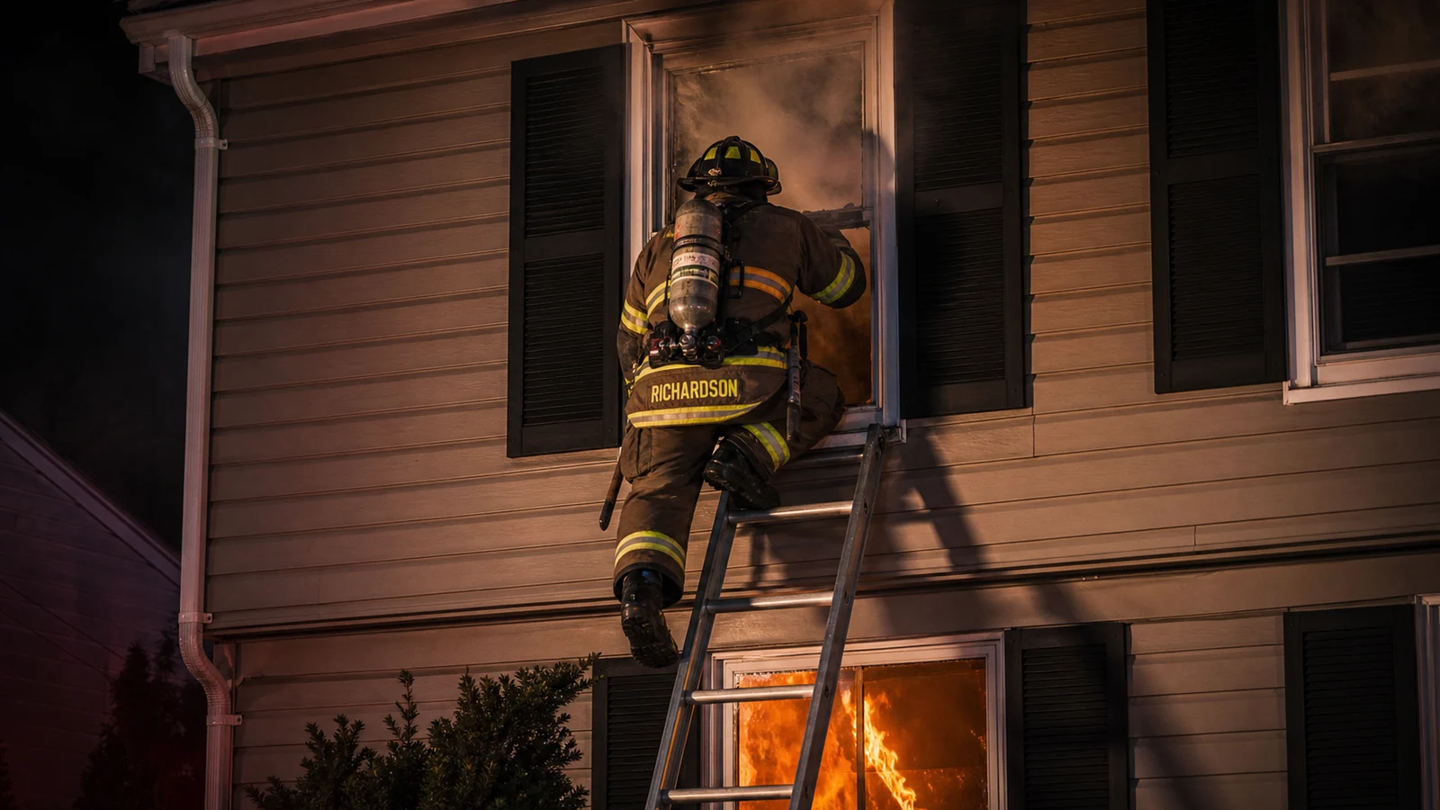
Cardiac Arrest Response for Firefighter-Medics: High-Performance CPR, Pit Crew Model & ROSC
Cardiac arrest is the highest-acuity call in EMS and one of the most common life-threatening emergencies fire companies respond to. Out-of-hospital cardiac arrest (OHCA) survival rates remain stubbornly low nationally — around 10% for all-cause OHCA — but departments with optimized protocols consistently achieve survival rates 2–3 times higher. The difference is almost entirely in how well the team executes the basics: high-quality CPR, early defibrillation, and a coordinated pit crew model. This guide covers the evidence-based cardiac arrest response for fire-based EMS.
Jump to:Chain of survival · High-quality CPR: what it actually means · Pit crew model · Airway management · Medications · ROSC and post-cardiac arrest care · Termination of resuscitation · Refractory arrest · FAQ
Chain of Survival: Every Link Matters
The AHA chain of survival for out-of-hospital cardiac arrest has six links. Each link that is weak reduces survival probability independently of how well the others are performed:
- Recognition and activation — Bystander recognizes cardiac arrest and calls 911 within 1 minute
- Early CPR — Bystander CPR before EMS arrival. Bystander CPR doubles to triples survival. This is the link most impacted by community education.
- Rapid defibrillation — AED application within 3–5 minutes of collapse for shockable rhythms (VF/pVT). Survival drops 7–10% for every minute without defibrillation.
- Advanced resuscitation — Fire/EMS ACLS interventions: high-quality CPR, advanced airway, medications
- Post-cardiac arrest care — Targeted temperature management, coronary intervention, ICU management
- Recovery — Rehabilitation and long-term support
Your job is links 3 and 4. Fire companies arriving on scene control the quality of defibrillation timing, CPR quality, and ACLS management. Everything after link 4 depends on what the field team delivers. The data is clear: high-performance CPR and early defibrillation are the interventions with the strongest evidence for survival improvement.
High-Quality CPR: What It Actually Means
The term "high-quality CPR" has specific, measurable criteria that are consistently associated with improved ROSC and neurologically intact survival:
| Parameter | Target | Common failure |
|---|---|---|
| Compression rate | 100–120 per minute | Too fast (>120) reduces fill time; too slow reduces perfusion pressure |
| Compression depth | At least 2 inches (5 cm), no more than 2.4 inches (6 cm) | Shallow compressions (most common failure); excessive depth causes injury |
| Full recoil | Chest returns to full resting position between every compression | Leaning on the chest — prevents ventricular filling |
| CCF (chest compression fraction) | ≥60%; target ≥80% | Pauses for pulse checks, airway management, rhythm analysis, movement |
| Pause duration | Pre-shock pause <10 seconds; post-shock pause immediate resumption | Long pauses for rhythm analysis or intubation |
| Ventilation rate | 1 breath per 6 seconds (10/min) with advanced airway | Hyperventilation — dramatically worsens outcome by increasing intrathoracic pressure |
Hyperventilation kills. Over-ventilation during CPR increases intrathoracic pressure, reduces venous return, and dramatically worsens coronary perfusion pressure. One breath every 6 seconds with an advanced airway in place. Not more. Monitor for visible chest rise — not for a rate.
Compressor fatigue
CPR quality degrades significantly after 2 minutes of continuous compressions, even in trained rescuers who are not aware of the decline. Rotate compressors every 2 minutes at minimum, timed with rhythm checks. The pit crew model structures this rotation so it happens automatically.
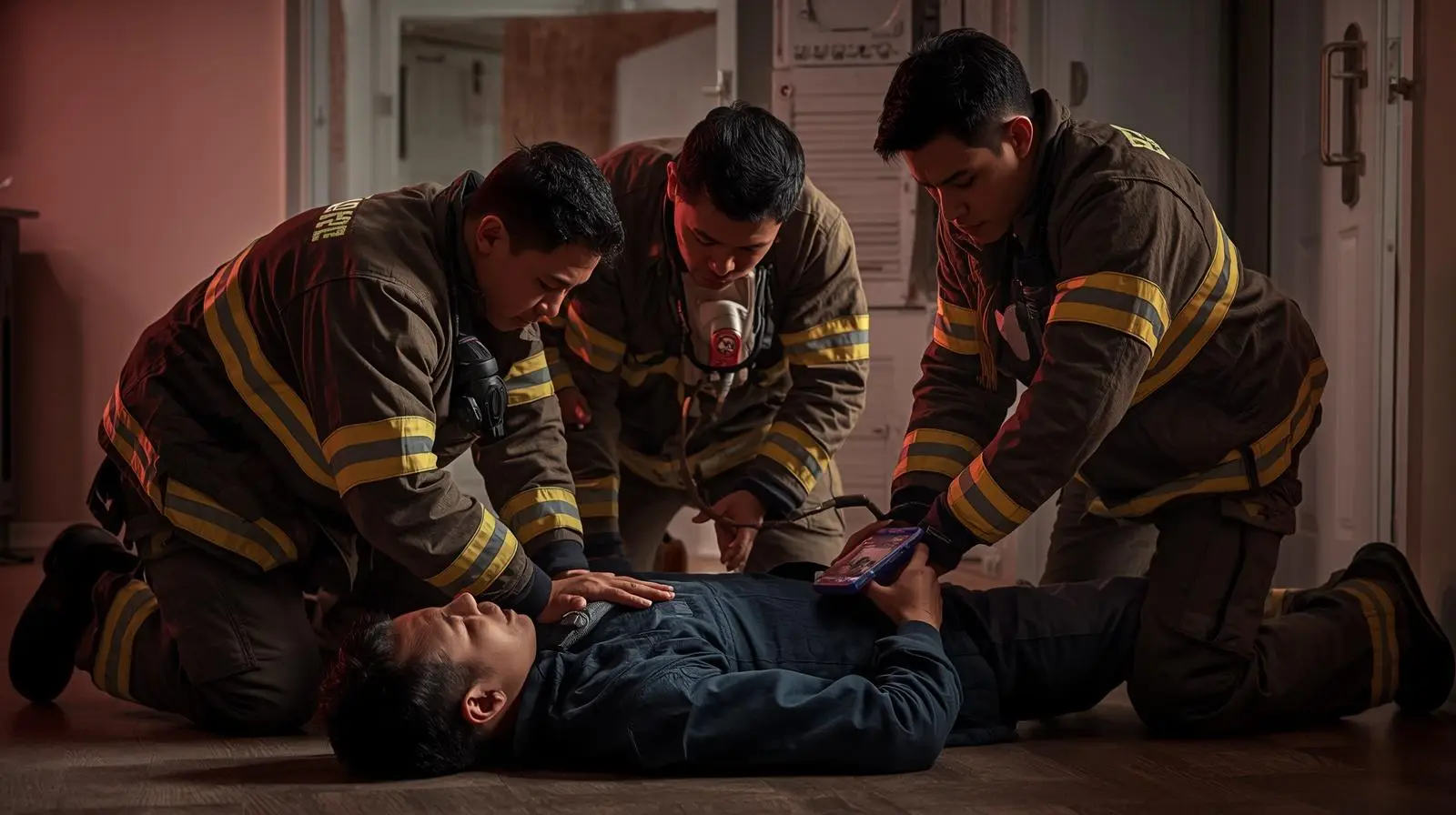
The Pit Crew CPR Model
The pit crew model assigns specific roles to each arriving crew member, so that compressions never stop while roles transition. The model eliminates the chaos of unstructured cardiac arrest management and ensures every intervention happens in parallel, not sequentially.
Standard 4-person pit crew assignments
| Role | Primary task | Secondary task |
|---|---|---|
| Compressor 1 (C1) | High-quality chest compressions from the patient's right side | Rotates to C2 on 2-minute cycle; calls fatigue early |
| Compressor 2 (C2) | Ready to rotate in; manages AED/monitor leads | Applies monitor pads; manages rhythm analysis during pulse check |
| Airway (AW) | BVM ventilation at head; monitors chest rise | Prepares for advanced airway when directed; suctions as needed |
| Team leader (TL) | Directs the resuscitation; calls rhythm checks; communicates with hospital | Documents time of collapse, interventions, medications; manages scene |
Rotation protocol (2-minute cycle)
The goal of the pit crew model is a CCF of 80%+. Every second the chest is not being compressed is a second without coronary perfusion pressure. Structure every transition to minimize that time.
Airway Management in Cardiac Arrest
Airway management in cardiac arrest has evolved significantly. The old model of early intubation as a priority has been replaced by an evidence-based approach that prioritizes compressions over airway interventions.
BVM first
For the first several minutes of resuscitation, BVM ventilation with a good mask seal is entirely adequate. Compressions should not be interrupted to intubate. If BVM is providing visible chest rise, continue with BVM until the compressor rotation provides a natural window for airway upgrade.
Advanced airway timing
Place an advanced airway (supraglottic device or endotracheal tube) during a compressor rotation transition, not during a compression cycle. The advanced airway should not interrupt CPR. Once an advanced airway is in place, compressions are continuous and ventilation is asynchronous at 1 breath per 6 seconds.
Supraglottic vs ETT
Recent evidence (AIRWAYS-2, PART trials) suggests supraglottic airways (King LT, iGel) achieve equivalent or better neurologic outcomes compared to ETT in out-of-hospital cardiac arrest, with faster placement and fewer interruptions. Know your protocol — many systems have moved to supraglottic as the primary advanced airway in OHCA.
Medications in Cardiac Arrest
| Medication | Indication | Dose | Evidence |
|---|---|---|---|
| Epinephrine 1:10,000 | All cardiac arrest rhythms | 1 mg IV/IO every 3–5 min | Improves ROSC; unclear effect on neurologic outcome. Give early in non-shockable rhythms. |
| Amiodarone | Refractory VF/pVT after 3 shocks | 300 mg IV/IO first dose; 150 mg second dose | Improves ROSC and survival to hospital admission in shock-refractory VF/pVT |
| Lidocaine | Refractory VF/pVT (alternative to amiodarone) | 1–1.5 mg/kg IV/IO | Similar to amiodarone in ALIVE trial; use per protocol |
| Sodium bicarbonate | Known hyperkalemia, TCA overdose, prolonged arrest | 1 mEq/kg IV/IO | Routine use not recommended; protocol-specific indications only |
| Calcium chloride | Hyperkalemia, hypocalcemia, calcium channel blocker OD | 1 g IV/IO (10% solution) | Protocol-specific; not routine in all arrests |
IO access is equivalent to IV in cardiac arrest. If IV access cannot be established quickly, intraosseous access provides reliable delivery of all cardiac arrest medications. Do not delay medication administration attempting to establish IV when IO can be placed in seconds.
ROSC and Post-Cardiac Arrest Care
Return of spontaneous circulation (ROSC) is not the end of the resuscitation — it is the transition to a new phase. Post-ROSC management in the field directly impacts neurologic outcome:
- Avoid hyperoxia. Once ROSC is achieved, titrate oxygen to SpO2 94–98%. Hyperoxia (SpO2 >98% with high-flow O2) is associated with worse neurologic outcomes after resuscitation.
- Avoid hypotension. Target a systolic BP ≥90 mmHg post-ROSC. Hypotension after ROSC is associated with secondary brain injury. Fluid bolus and vasopressors per protocol if BP is not maintained.
- 12-lead ECG immediately. STEMI after ROSC requires urgent cardiac catheterization. Recognize and communicate STEMI to the receiving facility before arrival so the cath lab can be activated en route.
- Temperature management. Avoid hyperthermia (>37.5°C). Targeted temperature management (TTM) at 32–36°C may be initiated in the field per protocol in some systems.
- Prevent re-arrest. Establish good IV access, maintain airway, monitor rhythm continuously. Re-arrest after ROSC is common and must be anticipated.
Termination of Resuscitation
Not every cardiac arrest should be resuscitated until hospital arrival. Evidence-based termination of resuscitation (TOR) rules help identify patients who are unlikely to survive transport and may benefit from field termination, which also improves safety by preventing high-speed transport with ongoing CPR.
The BLS TOR rule (Verbeek rule): consider termination if ALL three are true:
- Arrest not witnessed by EMS
- No AED shock delivered at any point
- No ROSC achieved after full ALS resuscitation
Always follow your local medical director's protocol and online medical control requirements for field termination. TOR rules are decision supports, not substitutes for physician oversight.
Refractory Cardiac Arrest
Refractory VF/pVT that does not respond to standard ACLS represents a growing area of advanced intervention. Options that some systems are implementing:
- Double sequential defibrillation (DSED): Two defibrillators applied simultaneously at different pad positions. Growing evidence from the DOSE-VF trial supports improved ROSC in refractory VF.
- ECPR (extracorporeal CPR): Venoarterial ECMO initiated in refractory arrest in appropriate candidates. Requires transport to an ECMO-capable center with rapid identification of candidates. EMS role: maintain perfusion to the hospital and communicate candidacy criteria.
- Point-of-care ultrasound (POCUS): Identifying and treating reversible causes (tamponade, pneumothorax, massive PE) that standard ACLS does not address.
Frequently Asked Questions
What is the target compression rate for CPR?
100–120 compressions per minute. Rates below 100 are inadequate to maintain coronary perfusion pressure. Rates above 120 reduce ventricular fill time and are associated with worse outcomes. Use a metronome, feedback device, or compression-assist technology to maintain the target rate.
What does CCF mean in cardiac arrest?
Chest compression fraction (CCF) is the percentage of time during a resuscitation that chest compressions are actually being performed. Target is ≥60%, with high-performance systems targeting ≥80%. Every pause in compressions — for pulse checks, intubation, rhythm analysis, or movement — reduces CCF and cardiac output.
When should epinephrine be given in cardiac arrest?
For non-shockable rhythms (PEA/asystole): as soon as IV/IO access is established, give epinephrine 1 mg and repeat every 3–5 minutes. For shockable rhythms (VF/pVT): after the second or third shock per most protocols, then every 3–5 minutes. Earlier epinephrine in non-shockable rhythms is associated with improved ROSC.
What is the pit crew model in EMS?
The pit crew model assigns specific, pre-defined roles to each arriving rescuer in a cardiac arrest: compressor 1, compressor 2 (who rotates on a 2-minute cycle), airway manager, and team leader. The model ensures compressions are continuous through transitions, every intervention is assigned to a specific person, and no one is improvising their role on arrival.


Comments 0
No comments yet. Be the first to share your thoughts!
Leave a Comment