

A patient who has been pinned under structural debris for three hours can be alert, conversational, and tell you their pain is manageable. Their blood pressure may be acceptable. Their pulse oximetry looks reasonable. Everything about them suggests someone who is injured but stable. Thirty minutes after extrication, they can be in cardiac arrest.
Crush syndrome — the systemic consequence of prolonged compression of large muscle groups — is one of the most counterintuitive medical emergencies that EMS and rescue personnel encounter. The compression itself is injuring the tissue continuously, but the most dangerous phase begins not when the weight is applied but when it is removed. The protocols that manage crush syndrome exist to bridge the gap between extrication and definitive hospital care in a way that reduces the probability of that deterioration — but the window for effective intervention is finite and starts before the patient is freed.
In this article:
- The mechanism: what compression does to muscle tissue
- The smiling patient: why stable presentation is misleading
- Reperfusion injury: why releasing the compression triggers the crisis
- Myoglobin and the kidneys
- Cardiac complications: hyperkalemia and arrhythmia
- The fluid protocol: why it starts before extrication
- Signs and symptoms EMS crews recognize
- Crush syndrome vs. compartment syndrome
The Mechanism: What Compression Does to Muscle Tissue
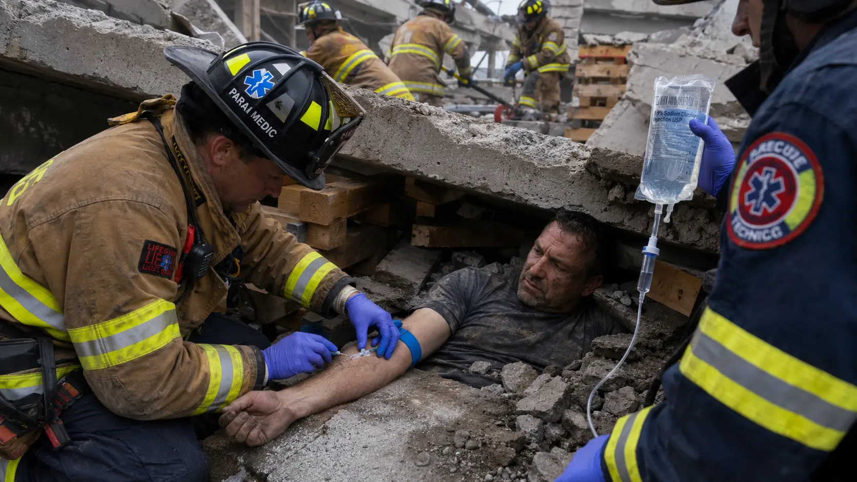
Skeletal muscle cells require a continuous blood supply to survive. When a muscle group is compressed by overlying weight — collapsed building debris, a vehicle dashboard, industrial equipment — the compression occludes blood vessels within the compressed tissue. The cells below the compression threshold continue their metabolic processes but without oxygen delivery or waste removal. Within 30 to 60 minutes, this ischemia begins producing irreversible cell death — rhabdomyolysis, the breakdown of skeletal muscle.
As muscle cells die, their contents are released. The most clinically significant component is myoglobin — the protein that stores oxygen in muscle tissue. Myoglobin is large, relatively insoluble at physiological pH, and toxic to kidney tubule cells at elevated concentrations. A person with significant rhabdomyolysis has circulating myoglobin levels that their kidneys must filter, and at high concentrations, the myoglobin precipitates in the tubules and causes acute kidney injury.
Two other cellular contents released in rhabdomyolysis are potassium and phosphate. Intracellular potassium concentration is approximately 140 mEq/L — roughly 35 times higher than normal serum potassium. When massive muscle cell death releases this potassium into the bloodstream simultaneously, serum potassium levels can rise to levels that produce life-threatening cardiac arrhythmias within minutes.
The Smiling Patient: Why Stable Presentation Is Misleading
The smiling patient is a clinical phenomenon that has been described repeatedly in earthquake mass casualty events, industrial accidents, and vehicle entrapments involving prolonged compression. The patient is alert, oriented, sometimes cheerful, and reports pain that is tolerable. Vital signs may be near-normal or only mildly abnormal.
The physiological explanation for this stability: while compressed, the injured extremities are effectively isolated from systemic circulation. The massive quantity of cellular breakdown products — myoglobin, potassium, lactic acid, inflammatory mediators — is trapped in the compressed tissue compartment. The patient's systemic circulation is not yet dealing with the full load of injury byproducts. The compression that is causing the injury is also, temporarily, containing the consequences of that injury.
This is the dangerous part. The rescue crew sees a patient who looks manageable. The temptation is to move quickly — get the weight off, get them out, get them to the hospital. Moving quickly without fluid loading first is the sequence that produces sudden deterioration. The moment compression releases, the trapped material floods into systemic circulation and the clinical picture changes radically.
Reperfusion Injury: Why Releasing the Compression Triggers the Crisis
Reperfusion injury — the paradoxical worsening of tissue damage when blood flow is restored — is a phenomenon observed in cardiac intervention, stroke treatment, and limb replantation as well as in crush syndrome. When blood flow is restored to ischemic tissue, the sudden availability of oxygen produces a surge of reactive oxygen species — free radicals that cause additional cell damage beyond what the original ischemia produced.
In crush syndrome, reperfusion has an additional mechanism: the restoration of blood flow to the compressed compartment flushes the accumulated cellular breakdown products into systemic circulation simultaneously. The kidneys, which had been receiving relatively normal blood chemistry while the compression was in place, are suddenly presented with a large bolus of myoglobin, potassium, phosphate, and inflammatory mediators that they must process at the same time that the reperfusion injury is damaging the previously compressed tissue further.
The clinical timeline: extrication occurs, the patient looks roughly similar to their pre-extrication state for some minutes, then blood pressure drops as fluid shifts into the damaged tissue (third spacing), potassium rises to arrhythmia-producing levels, and urine output drops as the kidneys begin to fail. Without aggressive fluid therapy that began before extrication, this timeline can end in cardiac arrest before hospital arrival.
Myoglobin and the Kidneys
Myoglobin becomes toxic to the kidneys at elevated serum concentrations through a specific mechanism: in the acidic environment of damaged kidney tubules, myoglobin precipitates and physically obstructs the tubular lumen. It also produces a direct toxic effect on tubule cells through oxidative damage. The result is acute tubular necrosis — the death of the filtering cells in the kidney tubules — and progressive renal failure.
The clinical sign that EMS can observe in the field is myoglobinuria: myoglobin appearing in the urine, producing a characteristic dark brown, "cola-colored" or "tea-colored" appearance. Any crush patient who produces urine in the field that is dark rather than yellow has significant circulating myoglobin and is at high risk for acute kidney injury. This finding should be communicated immediately to the receiving hospital to ensure dialysis capability is available.
The renal protective intervention is aggressive fluid diuresis — maintaining high urine flow to flush myoglobin through the tubules before it precipitates. This is why the fluid protocol is so important and why it must begin before extrication rather than waiting until hospital arrival.
Cardiac Complications: Hyperkalemia and Arrhythmia
The release of intracellular potassium from destroyed muscle cells into systemic circulation produces hyperkalemia — elevated serum potassium. Normal serum potassium is 3.5 to 5.0 mEq/L. Cardiac arrhythmias begin to appear at levels above 6.0 mEq/L. Ventricular fibrillation becomes likely above 8.0 mEq/L. In severe crush syndrome with extensive muscle involvement, potassium release can exceed the capacity of any intervention to contain, and cardiac arrest from hyperkalemia is the terminal event.
EKG changes in hyperkalemia follow a progression: peaked T-waves appear first, followed by prolonged PR interval and widened QRS complex, followed by a sine-wave pattern that immediately precedes ventricular fibrillation. A crush patient with a widening QRS on the monitor is in immediate cardiac jeopardy. Treatment in the field is limited to calcium gluconate (which stabilizes the cardiac membrane without reducing potassium levels) and sodium bicarbonate (which temporarily shifts potassium back into cells by alkalinizing the blood). Neither is a definitive treatment — both are bridges to hospital management.
The Fluid Protocol: Why It Starts Before Extrication
The single most important prehospital intervention in crush syndrome is establishing IV access and initiating aggressive fluid resuscitation before the compression is released. The target volume varies by protocol and by the estimated compression time, but a commonly cited target is 1 to 1.5 liters of normal saline per hour of compression, with a minimum of 1 liter delivered before extrication regardless of compression duration.
The rationale is straightforward: the fluid serves two purposes simultaneously. It pre-loads the vascular system to compensate for the fluid shifts and third spacing that will occur on reperfusion, preventing the hypotensive crash. And it prepares the kidneys for the myoglobin load by establishing high urine flow rates before myoglobin enters circulation in bulk — flushing the tubules continuously rather than allowing myoglobin to accumulate and precipitate.
In mass casualty events like earthquake responses, IV access and fluid loading of every confirmed or suspected crush patient before extrication is the protocol that has been associated with significantly lower rates of renal failure and mortality compared to extrication-first approaches. The 1988 Armenian earthquake response, the 1999 Marmara earthquake in Turkey, and the 2010 Haiti earthquake all produced data sets on crush syndrome management at scale that inform current protocols.
Signs and Symptoms EMS Crews Recognize
- ✗Prolonged compression history — any patient compressed for more than 1 hour with a large muscle group involved (thigh, both legs, torso) should be managed as a crush syndrome patient regardless of current presentation.
- ✗Swollen, firm extremities — compartment swelling visible even before extrication indicates significant edema and cellular destruction.
- ✗Dark, cola-colored urine — myoglobinuria; immediate indicator of significant rhabdomyolysis.
- ✗Peaked T-waves or QRS widening on EKG — hyperkalemia; immediate cardiac risk.
- ✗Rapid hemodynamic deterioration post-extrication — blood pressure drop immediately after release is the reperfusion flush; IV fluids running before this point significantly blunt the magnitude.
- ✓Establish IV access before extrication — even if the patient looks stable. Especially if the patient looks stable.
- ✓Run fluid aggressively — target 1L before release at minimum, continue running post-extrication.
- ✓Early hospital notification — receiving facility needs to have nephrology and dialysis capability ready.
Crush Syndrome vs. Compartment Syndrome
These two conditions involve related mechanisms but are clinically distinct. Compartment syndrome occurs when pressure within a closed muscle compartment — bounded by the fascia — rises to a level that occludes perfusion, typically from swelling after trauma. It can occur in limbs that were not crushed but were fractured or severely contused, and it develops over hours to days rather than being present at the time of extrication. The defining symptom is pain with passive stretch of the muscles in the affected compartment, and the treatment is surgical fasciotomy to decompress the compartment.
Crush syndrome is the systemic manifestation of massive rhabdomyolysis from prolonged compression — it is a whole-body crisis driven by the volume of cellular breakdown products, not a local compartment pressure problem. A crush patient may also develop compartment syndrome in the affected extremities after extrication as the reperfused tissue swells, which adds a surgical indication on top of the medical management already required. In significant crush incidents, compartment syndrome in the affected limbs is expected rather than exceptional.







Comments 0
No comments yet. Be the first to share your thoughts!
Leave a Comment