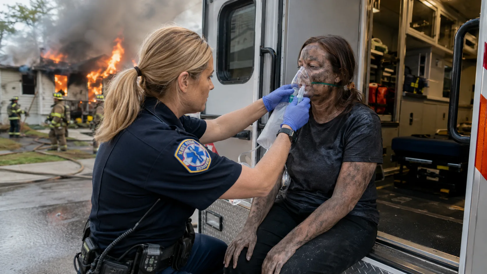

Smoke inhalation is the leading cause of death in residential structure fires — responsible for roughly 75 percent of fire fatalities. But smoke inhalation is not a single injury. It is a collection of three distinct injury mechanisms that operate simultaneously and at different timescales, which is why people who walked out of a fire feeling relatively well have died in the hospital 24 to 48 hours later, and why "I feel okay, I don't need to be checked out" is a statement that emergency medicine professionals consistently argue against.
Understanding what smoke actually does — the heat injury, the toxic gas injury, and the particle injury — explains both the symptoms and the reasons some of them are delayed, which symptoms warrant immediate emergency care, and what the treatment timeline looks like.
In this article:
- The three injury mechanisms
- Carbon monoxide and cyanide: the toxic gas injuries
- Delayed symptoms: why feeling okay is not a clearance
- Symptoms by severity: what warrants emergency care
- Children and the elderly: lower thresholds for concern
- What the ER evaluates and how
- Treatment: oxygen, hyperbaric, and cyanide antidote
- Recovery timeline and long-term effects
The Three Injury Mechanisms
1. Thermal injury
Hot gases and superheated air can burn the upper airway — the mouth, throat, and trachea — in ways that do not feel like what most people picture as a burn. The upper airway has less pain sensitivity than skin, and swelling from thermal injury develops over hours rather than immediately. A person who breathed hot air during a fire may feel only mild throat irritation at the scene. Six to twelve hours later, the swelling has developed to the point where the airway is significantly narrowed. This is the specific scenario that makes "I feel fine" dangerous — upper airway thermal injury can progress to complete airway obstruction in a person who was walking and talking hours earlier.
The lower respiratory tract — the lungs — is less susceptible to direct thermal injury because the upper airway acts as a heat exchanger, cooling air significantly before it reaches the lung tissue. Steam is an exception: steam carries substantially more heat energy per volume than dry hot air and can cause lower airway thermal injury at temperatures that dry air would not. Burns involving steam, pressurized hot liquids, or explosion in an enclosed space should be treated as having potential lower airway involvement regardless of surface presentation.
2. Toxic gas injury
Smoke contains dozens of toxic compounds generated by the incomplete combustion of building materials, furnishings, and contents. Carbon monoxide and hydrogen cyanide are the most immediately life-threatening. Both impair oxygen delivery to tissues — CO by binding hemoglobin, cyanide by blocking cellular oxygen utilization — and both produce effects that are not visible or obviously traceable to smoke without laboratory testing. Other toxic gases present in structure fire smoke include hydrogen chloride (from burning PVC), acrolein (from burning plastics and wood), and phosgene (from certain chemical combustion reactions). Each has its own toxicological profile and contributes to the overall injury burden.
3. Particle deposition
Fine particulate matter in smoke — the same PM2.5 fraction that makes wildfire smoke hazardous — deposits in the lung tissue and triggers inflammatory responses. In the acute phase, this produces bronchospasm (the airways tighten, making breathing difficult) and increased mucus production. In the hours and days after significant exposure, the inflammatory response can produce pulmonary edema — fluid accumulation in the lungs — that worsens respiratory function progressively rather than improving. This is another mechanism behind delayed symptom onset.
Carbon Monoxide and Cyanide: The Toxic Gas Injuries
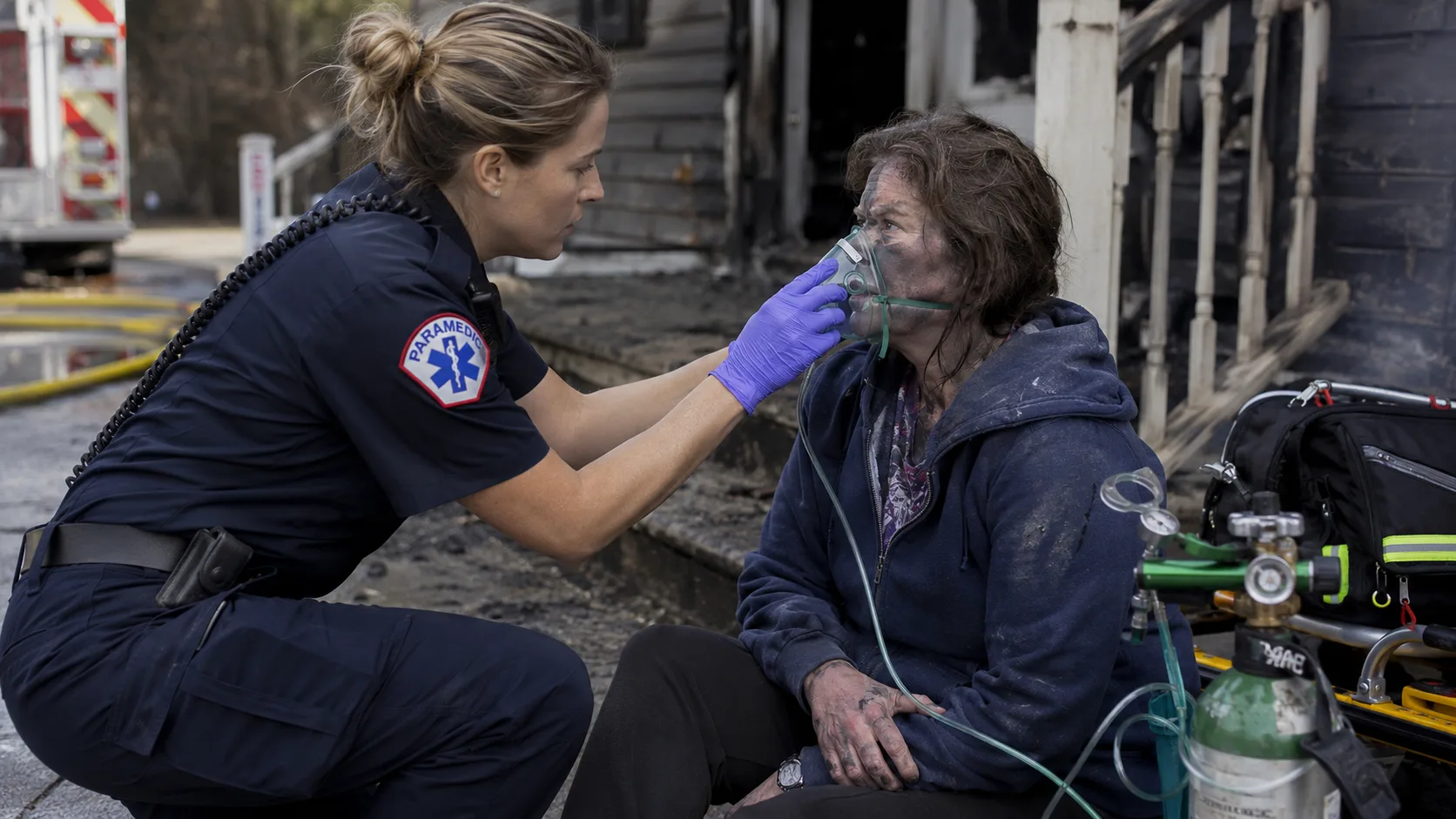
Carbon monoxide and hydrogen cyanide are both present in structure fire smoke, and both produce toxicity that is not visible and not necessarily symptomatic at the scene. CO poisoning is covered in detail in a separate article on this site; the key points relevant to smoke inhalation are that carboxyhemoglobin levels must be measured by blood test or pulse CO-oximetry to quantify exposure, and that levels can be deceptively low if significant time has passed between exposure and measurement (because CO begins to clear with fresh air exposure).
Hydrogen cyanide toxicity became a recognized component of structure fire smoke injury as building materials shifted toward synthetic content. Wood and natural fiber combustion produces primarily CO. Modern furniture, carpeting, and building materials containing nitrogen — nylon, polyurethane, acrylonitrile — produce cyanide compounds in combustion. The clinical presentation of cyanide toxicity overlaps significantly with CO poisoning: confusion, metabolic acidosis, cardiovascular instability. The differentiation matters because the treatments are different.
Emergency medicine now recognizes that treating only for CO in a smoke inhalation patient who is not improving may miss co-occurring cyanide toxicity. Hydroxocobalamin — a cyanide antidote — is carried by an increasing number of ALS (advanced life support) units and has been adopted into smoke inhalation protocols in many major fire departments' EMS systems. A patient who is not responding to high-flow oxygen after smoke exposure may have cyanide toxicity as a contributing factor.
Delayed Symptoms: Why Feeling Okay Is Not a Clearance
The medical literature on smoke inhalation is explicit on this point: feeling well at the scene is not a reliable indicator of injury severity. Three mechanisms produce delayed deterioration:
Upper airway swelling, as described above, develops over hours. A person with thermal injury to the upper airway who is breathing comfortably at the scene may be significantly compromised six hours later. The window for securing the airway electively — before it becomes an emergency intubation under pressure — is in the first hours after exposure. This is one of the primary reasons emergency physicians evaluate smoke exposure patients for airway injury even when they appear well.
Pulmonary edema from inflammatory particle deposition can develop 24 to 48 hours after exposure. The lungs appear to function normally initially; the inflammatory response produces fluid accumulation over time. Patients discharged from the emergency department as apparently stable have developed respiratory failure in this window. Extended observation or follow-up is appropriate for significant smoke exposure even without immediate symptoms.
CO poisoning neurological effects — cognitive impairment, memory deficits, personality changes — can develop days to weeks after apparently successful treatment. This syndrome, called delayed neurological sequelae, is more common after severe initial poisoning but can occur after moderate exposure. Hyperbaric oxygen therapy is thought to reduce the incidence of delayed neurological sequelae compared to normobaric oxygen treatment alone, which is part of the rationale for hyperbaric treatment in significant CO poisoning.
Symptoms by Severity: What Warrants Emergency Care
| Symptom | Significance | Action |
|---|---|---|
| Mild cough, mild throat irritation, mild headache | Common after any smoke exposure; may resolve with fresh air | Monitor — if symptoms persist or worsen, seek evaluation |
| Hoarse voice, barky cough, stridor (high-pitched breathing sound) | Suggests upper airway involvement — possible thermal injury with progressive swelling | Emergency evaluation immediately — airway injury can progress rapidly |
| Shortness of breath, wheezing | Bronchospasm or lower airway involvement; may indicate significant particle deposition | Emergency evaluation — respiratory status may worsen |
| Confusion, altered mental status | Suggests CO poisoning, cyanide toxicity, or significant hypoxia | Emergency evaluation immediately — impaired cognition may prevent self-report of worsening |
| Loss of consciousness, even brief | Indicates significant toxic exposure or hypoxia | Emergency evaluation immediately — do not defer |
| Soot around mouth and nose, singed nasal hair | External evidence of heat exposure to the upper airway | Emergency evaluation — airway assessment required even without symptoms |
| Persistent headache, nausea after fresh air exposure | Suggests CO poisoning | Emergency evaluation — CO level measurement needed |
Children and the Elderly: Lower Thresholds for Concern
Children have smaller airways that swell proportionally more significantly with the same degree of thermal injury — a degree of upper airway swelling that produces mild symptoms in an adult may produce significant obstruction in a child. Children also have higher respiratory rates, meaning they inhale more smoke per unit of body weight per minute than adults in the same smoke concentration. Both factors lower the threshold for symptom development and for serious injury at lower exposure levels.
Older adults have reduced respiratory reserve — the lungs have less capacity to compensate for injury, and pre-existing conditions (COPD, heart disease) lower the threshold at which smoke inhalation produces serious physiological consequences. A smoke exposure that a healthy 35-year-old manages without significant difficulty may produce severe respiratory compromise in a 75-year-old with pre-existing COPD. Any smoke exposure in an older adult with cardiopulmonary conditions warrants evaluation regardless of apparent symptom severity.
What the ER Evaluates and How
Emergency evaluation of a smoke inhalation patient includes several components that cannot be assessed without medical equipment:
- Carboxyhemoglobin level — measured by co-oximetry on a blood sample or by a non-invasive pulse CO-oximeter. Standard pulse oximeters cannot distinguish between oxyhemoglobin and carboxyhemoglobin — a patient with significant CO poisoning may have a normal-appearing standard pulse ox reading. This is a critical diagnostic limitation that is not widely understood outside of emergency medicine.
- Airway assessment — direct or video laryngoscopy to visualize the upper airway for evidence of thermal injury, edema, or soot deposition. This cannot be performed without equipment and is the standard of care for patients with any sign of potential airway involvement.
- Chest X-ray and/or CT — to assess for early pulmonary infiltrates, atelectasis, or other lower airway findings.
- Arterial blood gas — to assess oxygenation, ventilation, and metabolic acidosis (which suggests cyanide toxicity or significant CO poisoning).
- Lactic acid level — elevated lactic acid suggests cellular oxygen deprivation consistent with cyanide toxicity.
Treatment: Oxygen, Hyperbaric, and Cyanide Antidote
High-flow oxygen via non-rebreather mask is the first-line treatment for CO poisoning — 100% oxygen delivered at high flow rates accelerates CO elimination from hemoglobin approximately four times faster than breathing room air. This treatment should begin at the scene, not after transport.
Hyperbaric oxygen — 100% oxygen at 2 to 3 atmospheres of pressure in a pressurized chamber — accelerates CO elimination significantly faster than normobaric high-flow oxygen and is thought to reduce the incidence of delayed neurological sequelae. It is indicated for severe CO poisoning (loss of consciousness, neurological symptoms, significant carboxyhemoglobin levels, pregnancy, cardiac involvement). Not all hospitals have hyperbaric chambers; patients who meet criteria may be transferred to a facility with this capability.
Hydroxocobalamin (Cyanokit) is the cyanide antidote with the best evidence base for smoke inhalation management. It reacts with cyanide to form a non-toxic compound that is renally excreted. It is given empirically in patients with severe smoke inhalation and signs of cyanide toxicity (elevated lactate, hemodynamic instability not responding to oxygen) because waiting for laboratory confirmation of cyanide poisoning is clinically impractical.
Recovery Timeline and Long-Term Effects
Mild smoke inhalation — brief exposure, no symptoms beyond transient throat irritation, normal airway examination — typically resolves without lasting effects within a few days. Moderate to severe exposure with documented injury requires a longer recovery trajectory.
Upper airway injury typically resolves within 1 to 2 weeks without permanent changes if appropriately managed. Lower airway injury from particle deposition can produce a prolonged inflammatory phase — persistent cough, reduced exercise tolerance, increased airway reactivity — lasting weeks to months. Patients who develop reactive airways disease (bronchospasm) that was not present before the fire exposure may have new-onset asthma that persists long-term.
CO poisoning with delayed neurological sequelae — the cognitive and personality changes that can emerge days to weeks after the acute event — may take months to resolve and in severe cases may not fully resolve. Neuropsychological testing several weeks after significant CO poisoning can identify deficits that are not apparent on casual evaluation. Follow-up with a neurologist is appropriate for patients who experienced loss of consciousness or significant neurological symptoms during their CO poisoning.
Anyone who has had significant smoke inhalation should be monitored for the development of respiratory symptoms in the days and weeks after exposure, maintain follow-up with their primary care physician, and should not assume that feeling well shortly after the event represents complete resolution of the injury process.

Comments 0
No comments yet. Be the first to share your thoughts!
Leave a Comment